
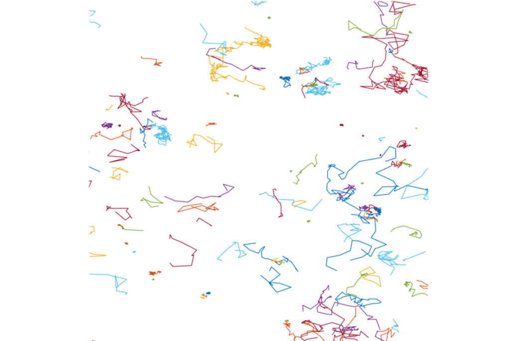
Single molecule techniques investigate the complex behaviors of biomolecules in vitro and in cellular environments, revealing their molecular structure, dynamics, interactions, and functions. By avoiding the need for averaging that is inherent in bulk measurements, these powerful methods provide nuanced insights into the diverse nature of molecular activities, enhancing our understanding of molecular heterogeneity.
Researchers

We are interested in understanding the underlying principals that govern the functions of intrinsically disordered proteins and why these proteins are so frequently associated with disease. To do this, we use single-molecule fluorescence and other biophysical techniques to study protein conformations and dynamics, protein-protein interactions, and protein-membrane interactions. We are interested in understanding the underlying principals that govern the functions of intrinsically disordered proteins and why these proteins are so frequently associated with disease. To do this, we use single-molecule fluorescence and other biophysical techniques to study protein conformations and dynamics, protein-protein interactions, and protein-membrane interactions.
Single Molecule Imaging

The overarching goal of Lakadamyali lab is to understand the mechanisms that regulate sub-cellular organization and the significance of this organization for organelle and cell function. To study the spatiotemporal organization of cells with unprecedented quantitative detail, we combine cell biological tools with new live-cell and super-resolution microscopy methods. Lakadamyali lab has developed novel live-cell and super-resolution microscopy methods with enhanced spatiotemporal resolution, high-throughput multi-color super-resolution microscopy methods, and advanced computational and quantitative tools for super-resolution microscopy. These tools enable us to address key biological questions including how microtubules and motor protein complexes regulate organelle transport and positioning under physiological and pathological conditions, how organelle positioning relates to organelle molecular identity and influences organelle function, and what is the cause/effect relationship between the nucleosome level folding of the chromatin fiber and cellular identity in differentiation, reprogramming and diseases.
Single Molecule Imaging

The goal of our research is to understand the cellular machinery responsible for powering cell movements and shaping the architecture of cells, tissues, and organs. Our discovery-based research focuses on the role of the cytoskeleton, molecular motors, and signaling pathways in powering cell migration, muscle contraction, and the transport of internal cell compartments. The pathways investigated in our laboratory are crucial for several normal and pathological processes, including: cell and tissue development, endocytosis, wound healing, immune response, cardiomyopathies, and metastases of tumors.
Single Molecule Imaging cryoEM

More than 60% of current drug targets are membrane proteins, which come in the form of enzymes, receptors, channels, and transporters. This underlines the biomedical relevance of our research into the function of membrane proteins and lipids.
Our research is highly interdisciplinary and collaborative. Our group members typically have backgrounds in fields such as physical chemistry, chemical biology, biochemistry, physics, and various engineering disciplines, and we collaborate with multiple different groups in our department, elsewhere on campus, or at nearby research institutions.
Specifically, we are interested in the function of lipid transporters (flippases) and how these can be modulated through photopharmacology, the structure and function of proteins involved in endocytosis (using techniques such as Cryo Electron Tomography and various fluorescence labeling, microscopy, and spectroscopy approaches), the function of intrinsically disordered proteins on membranes (using 2D NMR spectroscopy and various fluorescence techniques), all complemented with micromanipulation techniques and interpretation with thermodynamic and statistical mechanical models and simulations. A recent development in our lab has been to ask to what extent and by what mechanisms protein-protein liquid phase separation, referred to as LLPS, contributes to some of these phenomena.
Single Molecule Imaging cryoEM

Our laboratory is focused on understanding the dynamics of intracellular motility. The active transport of membrane-bound organelles such as mitochondria along the cytoskeleton is essential in most eukaryotic cells, but is especially important in neurons. Neurons are highly polarized cells, with axons that can extend up to one meter, making them uniquely dependent on motor-driven transport. We explore the molecular mechanisms that lead to the coordinated activity of molecular motors during long-distance transport. Mutations in motors or required activators are sufficient to cause neuro-developmental and/or neurodegenerative disease; thus we study transport deficits in the context of human diseases including ALS, Huntington’s disease and Parkinson’s disease.
Organelle quality control is also essential to maintain healthy neurons. We explore the dynamics of autophagy, mitophagy, and lysophagy in neurons, examining biogenesis, cargo capture, trafficking, and cargo degradation. Mutations in key genes including PINK1, Parkin, OPTN, TBK1, and LRRK2 disrupt autophagy in neurons, strongly implicating defects in these pathways in neurodegenerative diseases including ALS and Parkinson's.
We focus on mechanistic cell biology, with approaches including: live cell imaging of cytoskeletal and organelle dynamics in primary neurons and human iPSC-derived neurons, in vitro reconstitution assays using TIRF microscopy to obtain single molecule resolution, and analysis of cellular and mouse models of neurodegenerative disease. Our lab takes an inclusive and collaborative approach to science, working with exceptional labs at Penn and worldwide, through the Pennsylvania Muscle Institute, the RM1 Mitochondrial Group, and the ASAP Collaborative Science Network.
Our success is built on our diversity; we are committed to mentoring scientists from a broad array of backgrounds and to supporting and enhancing opportunities for women and under-represented minorities in science.
Single Molecule Imaging cryoET